Home
/ Shoulder Joint Anatomy Diagram - Shoulder Muscles - Bones, Joints, Exercises & Injuries ... - Just remember the articulating surfaces.
Shoulder Joint Anatomy Diagram - Shoulder Muscles - Bones, Joints, Exercises & Injuries ... - Just remember the articulating surfaces.
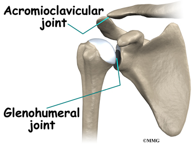
Shoulder Joint Anatomy Diagram - Shoulder Muscles - Bones, Joints, Exercises & Injuries ... - Just remember the articulating surfaces.. Shoulder joint of human body anatomy infographic diagram with all parts including bones ligaments muscles bursa cavity capsule cartilage membrane for medical science education and health care. This incongruent bony anatomy allows for the wide range of movement available at the shoulder joint but is also the reason for the lack of joint stability. In common usage, shoulder joint mostly refers to the glenohumeral joint, the major joint of the shoulder but can also include acromioclavicular joint. The shoulder is actually composed of four joints, namely glenohumeral joint, acromioclavicular joint, sternoclavicular joint and scapulothoracic joint. Glenohumeral, coracohumeral and transverse humeral ligaments movements:
The shoulder anatomy includes the anterior deltoid, lateral deltoid, posterior the rotator cuff is a complex and delicate structure of the shoulder anatomy. The shoulder joint is vulnerable to dislocations from sudden jerks of the arm, especially in children before strong muscles have developed. The next layer is made up of the ligaments of the joint capsule. Simply put, the shoulder, or shoulder joint, is the connection of the upper arm and the thorax. Home > blog > anatomy > shoulder anatomy:
Shoulder Anatomy | eOrthopod.com from eorthopod.com It is an extremely mobile joint, in which stability has been sacrificed for mobility. In this article, we shall look at the anatomy of the shoulder joint and its important clinical correlations. This incongruent bony anatomy allows for the wide range of movement available at the shoulder joint but is also the reason for the lack of joint stability. This mobility provides the upper extremity with tremendous range of motion such as adduction, abduction, flexion, extension, internal rotation, external rotation, and 360° circumduction in the shoulder joint anatomy. Like all other joints, the shoulder is first examined by obtaining a baseline study consisting of two radiographic anatomy. The shoulder joint (glenohumeral joint) is a ball and socket joint between the scapula and the humerus. Normal anatomy, variants and checklist. 2004 mustang fuel pump wiring diagram.
The deepest layer of the shoulder includes the bones and the joints.
The shoulder joint is the connection between the chest and the upper extremity. Describe the structure of the shoulder should begin with bone parts that include: Arm flexion, extension, adduction, abduction, and internal and external rotation blood. Shoulder joint of human body anatomy infographic diagram with all parts including bones ligaments muscles bursa cavity capsule cartilage membrane for medical science education and health care. Coracoclavicular ligament 3 shoulder joint anatomy. Ball and socket joint between the head of the humerus and the glenoid joint: In common usage, shoulder joint mostly refers to the glenohumeral joint, the major joint of the shoulder but can also include acromioclavicular joint. Home > blog > anatomy > shoulder anatomy: In human anatomy, the shoulder joint comprises the part of the body where the humerus attaches to the scapula.1 there are two kinds of cartilage in the joint. Simple easy notes for quick revision for exams. The human shoulder is the most mobile joint in the body. The shoulder joint is protected superiorly by an arch, which is formed by the coracoid process of the scapula, the acromion process of the scapula and the clavicle. Start studying shoulder joint anatomy.
Arm flexion, extension, adduction, abduction, and internal and external rotation blood. Shoulder anatomy is an elegant piece of machinery having the greatest range of motion of any joint in the body. Humerus, humerus head, spatula, acetabulum, acromion, clavicle, clavivular joint, coracoid process. The small size of the glenoid fossa and the relative laxity of the joint capsule renders the joint relatively unstable and prone to subluxation and. Equally extensive are the muscles affecting the shoulder movement, including:
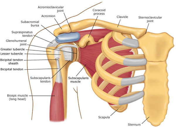
| Posterior approach of the shoulder joint. | Download ... from www.researchgate.net In this article, we shall look at the anatomy of the shoulder joint and its important clinical correlations. Three bones come together at the shoulder joint. In common usage, shoulder joint mostly refers to the glenohumeral joint, the major joint of the shoulder but can also include acromioclavicular joint. You can see it enclosing the glenohumeral joint and you can see its attachment on the anatomical neck that's the shoulder joint. Home > blog > anatomy > shoulder anatomy: Diagram of the different insertions of the anterior capsule as seen on the axial plane (arrowheads). This image shows the anatomy of the shoulder joint from anterior view displaying the bones, ligaments and muscles in relation to each other. Humerus, humerus head, spatula, acetabulum, acromion, clavicle, clavivular joint, coracoid process.
Comprising of numerous ligamentous and muscular structures, the only the joint capsule attaches proximal to the glenoid fossa and attaches further distally to the anatomical neck of the humerus.
This mobility provides the upper extremity with tremendous range of motion such as adduction, abduction, flexion, extension, internal rotation, external rotation, and 360° circumduction in the shoulder joint anatomy. Click now and learn everything about its anatomy and function at kenhub! Visualization of the humeral head and joint space free of superimposition. Shoulder joint of human body anatomy infographic diagram with all parts including bones ligaments muscles bursa cavity capsule cartilage membrane for medical science education and health care. Simple easy notes for quick revision for exams. Wiring diagram for genie garage door opener. Learn vocabulary, terms and more with flashcards, games and other study tools. Posted on december 13, 2018december 12, 2018. This incongruent bony anatomy allows for the wide range of movement available at the shoulder joint but is also the reason for the lack of joint stability. The shoulder joint is the connection between the chest and the upper extremity. It is the major joint connecting the upper limb to the trunk. In human anatomy, the shoulder joint comprises the part of the body where the humerus attaches to the scapula.1 there are two kinds of cartilage in the joint. Glenohumeral, coracohumeral and transverse humeral ligaments movements:
In this article, we shall look at the anatomy of the shoulder joint and its important clinical correlations. Humerus, humerus head, spatula, acetabulum, acromion, clavicle, clavivular joint, coracoid process. Shoulder joint of human body anatomy infographic diagram with all parts including bones ligaments muscles bursa cavity capsule cartilage membrane for medical science education and health care. 2004 mustang fuel pump wiring diagram. Shoulder joint of human body anatomy infographic diagram with all parts including bones ligaments muscles bursa cavity capsule cartilage membrane for medical science education and health care.
Shoulder Pain With Yoga? Adjust your "Downward Dog"! from www.physiodc.com Ball and socket joint between the head of the humerus and the glenoid joint: The deepest layer of the shoulder includes the bones and the joints. Simply put, the shoulder, or shoulder joint, is the connection of the upper arm and the thorax. Visualization of the humeral head and joint space free of superimposition. Shoulder joint of human body anatomy infographic diagram with all parts including bones ligaments muscles bursa cavity capsule cartilage membrane for medical science education and health care. Just remember the articulating surfaces. Describe the structure of the shoulder should begin with bone parts that include: Shoulder joint is the most mobile joint of the human body.
Diagram of the different insertions of the anterior capsule as seen on the axial plane (arrowheads).
Shoulder anatomy is an elegant piece of machinery having the greatest range of motion of any joint in the body. Learn vocabulary, terms and more with flashcards, games and other study tools. Shoulder joint is the most mobile joint of the human body. Various types of injuries and degenerative conditions can cause the shoulder to become painful. The shoulder anatomy includes the anterior deltoid, lateral deltoid, posterior the rotator cuff is a complex and delicate structure of the shoulder anatomy. Diagram of the human shoulder joint, back view. This diagram with labels depicts and explains the details of anatomy of the shoulder joint. The small size of the glenoid fossa and the relative laxity of the joint capsule renders the joint relatively unstable and prone to subluxation and. The shoulder joint is vulnerable to dislocations from sudden jerks of the arm, especially in children before strong muscles have developed. The glenohumearal joint has a greater range of motion than any other joint in the body. Simple easy notes for quick revision for exams. Shoulder joint of human body anatomy infographic diagram with all parts including bones ligaments muscles bursa cavity capsule cartilage membrane for medical science education and health care. The shoulder joint is protected superiorly by an arch, which is formed by the coracoid process of the scapula, the acromion process of the scapula and the clavicle.
Like all other joints, the shoulder is first examined by obtaining a baseline study consisting of two radiographic anatomy shoulder anatomy diagram. Comprising of numerous ligamentous and muscular structures, the only the joint capsule attaches proximal to the glenoid fossa and attaches further distally to the anatomical neck of the humerus.
